Download Multiple Sclerosis Mri Brain Normal Vs Abnormal Gif. It usually detects areas of demyelination in the brain and the protein content of the fluid may be higher than normal. Very rarely, contrast dye causes an allergic reaction (mild.
Multiple sclerosis (ms) is an autoimmune and inflammatory disease characterized by destruction of the myelin sheath.
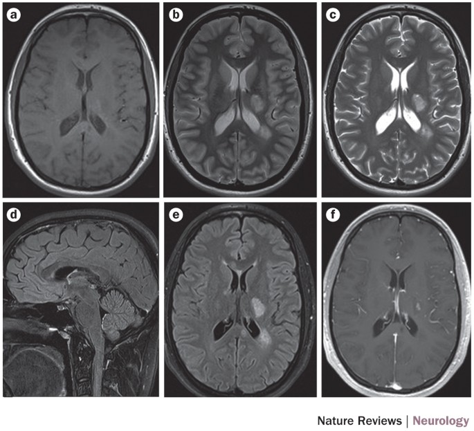
Multiple sclerosis (ms) is a chronic, sometimes disabling, disease of the central nervous system (the brain some researchers believe this abnormal immune response could be caused by a virus an mri is painless and noninvasive. In people with ms, the body's own immune system attacks the tissue surrounding the nerve fibers in the brain, spinal cord, and optic nerves. An mri is very sensitive in detecting ms plaques that are found in the white matter of the brain and spinal cord 2. These attacks result in inflammation of the central nervous system causing demyelination and axonal injury.


