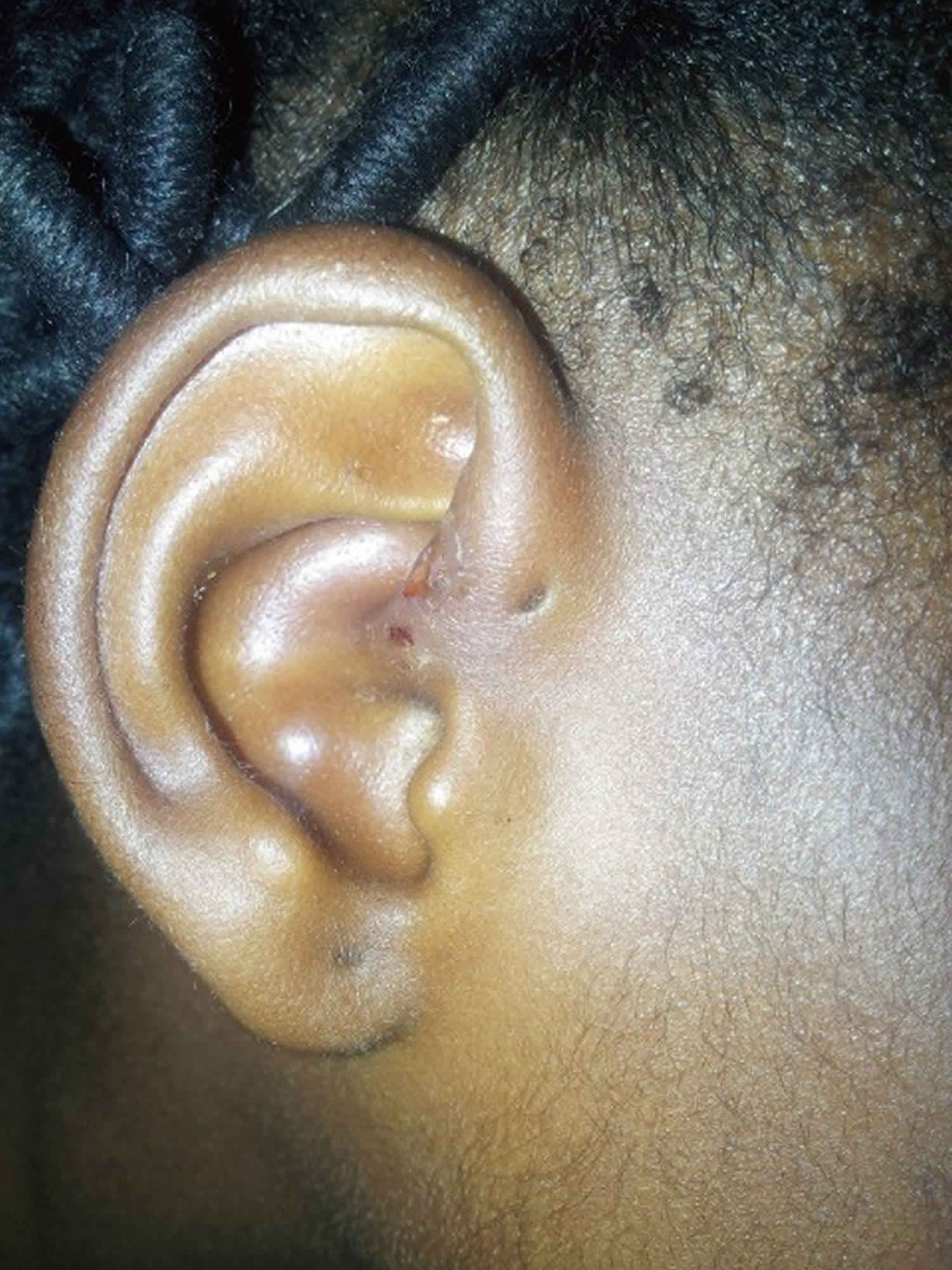
Download Drainage Preauricular Sinus Anatomy Pictures. A preauricular sinus is a common congenital malformation characterized by a nodule, dent or dimple located anywhere adjacent to the external ear. Preauricular sinuses are frequently noted on routine physical examination as small dells adjacent to the external ear, usually at the anterior margin of the ascending limb of the helix.

Preauricular sinuses and cysts result from developmental defects of the first and second branchial arches.4 occasionally a preauricular sinus or a cyst most preauricular sinuses are asymptomatic and remain untreated unless they become infected too often.6 preauricular cysts are treated with.
The tract may be of variable length, have a tortuous course, and exhibit extensive branching. Learn vocabulary, terms and more with flashcards, games and other study tools. Superior and inferior opthalmic veins drain the eye. Preauricular sinus is a common birth defect that may be seen during a routine exam of a newborn.


