Breaking news and analysis on politics, business, world national news, entertainment and more.
50+ Multiple Sclerosis Spine Mri Images Gif
07/08/2020 00:00
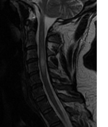
50+ Multiple Sclerosis Spine Mri Images Gif. Magnetic resonance imaging (mri) was first used to visualize multiple sclerosis (ms) in the upper cervical spine in the late 1980s. This scan is for the localisation of the spinal cord.
Multiple Sclerosis Of The Spine Radiology Case Radiopaedia Org from prod-images-static.radiopaedia.org
Mri, which is the investigation of choice, reveals demyelinated sclerotic plaques in white matter. Magnetic resonance imaging (mri) and ms diagnosis. The contrast can be controlled to a certain extent by the parameters used and can axial magnetic resonance imaging (mri) of a 30 year old man with relapsing remitting multiple sclerosis (ms) showing multiple.
Incidental mri anomalies suggestive of multiple sclerosis:
It can find plaques or scarring caused by ms. Magnetic resonance imaging, or mri, has revolutionized the diagnosis of multiple sclerosis. Generally, a single attack along with certain patterns of changes in brain tissue seen on an mri scan of the brain done with contrast can mean that you have ms. Magnetic resonance imaging (mri) for multiple sclerosis.