41+ Multiple Sclerosis Full Spine Mri Pics. Magnetic resonance imaging (mri) was first used to visualize multiple sclerosis (ms) in the upper cervical spine in the late 1980s. Multiple sclerosis (ms), also known as encephalomyelitis disseminata, is a demyelinating disease in which the insulating covers of nerve cells in the brain and spinal cord are damaged.
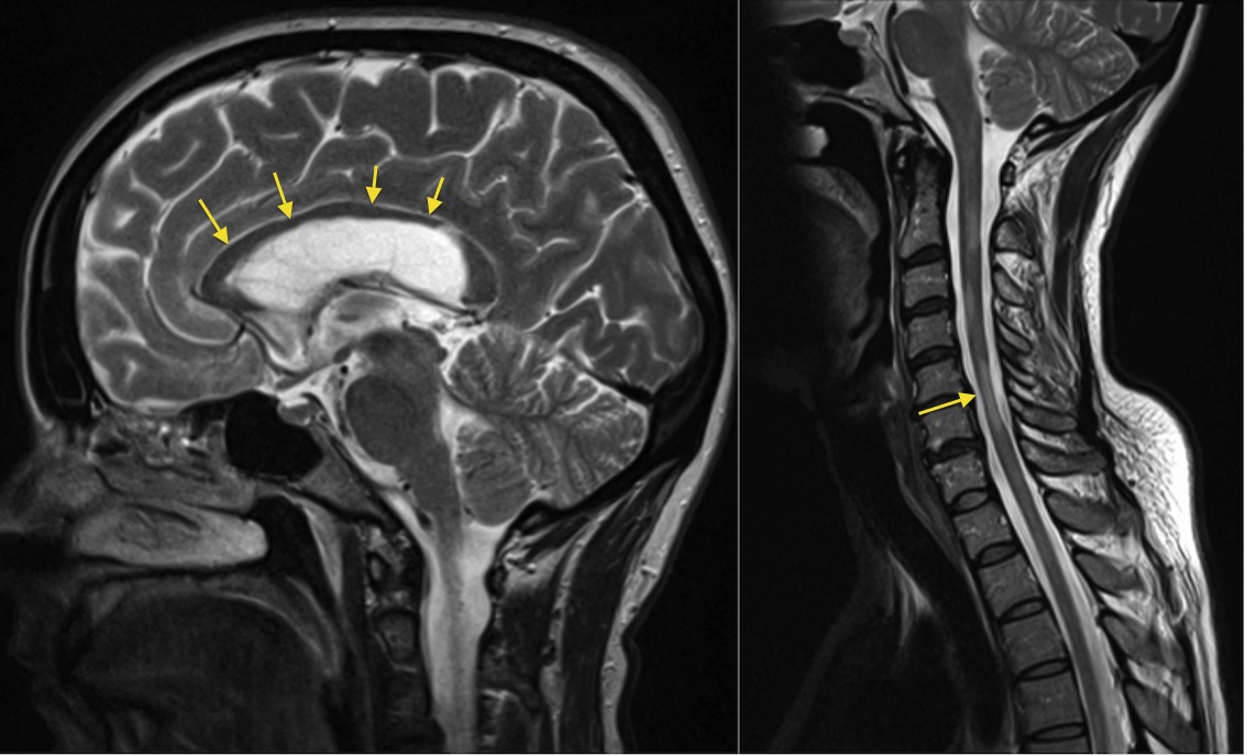
Mri, which is the investigation of choice, reveals demyelinated sclerotic plaques in white matter.
Peripheral nerves are not affected. And several of blood tests.4 the. This thesis explores abnormalities in the multiple sclerosis (ms) spinal cord and their relationship with physical disability through the use in summary, this thesis shows that ms spinal cord abnormalities may be visualised and quantified using high field mri, and are significantly associated with disability. Residents and fellows contest rules | international ophthalmologists contest rules.


