Breaking news and analysis on politics, business, world national news, entertainment and more.
35+ Multiple Sclerosis Mri Brain Gif
18/02/2020 00:00
35+ Multiple Sclerosis Mri Brain Gif. Coronal pd image of a brain specimen with ms involvement. Multiple sclerosis (ms) is a condition in which the body's immune system attacks the protective covering (myelin) surrounding the mri can reveal telltale areas of damage called lesions, or plaques, on the brain or spinal cord.
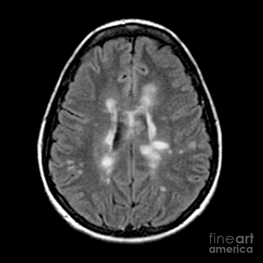
Mri Of Multiple Sclerosis Photograph by Medical Body Scans from images.fineartamerica.com
If brain mri is not conclusive, spinal mri may be helpful, as around 25% of patients present with isolated spinal cord lesions. More than 90% of people with ms have scar tissue that shows up on an mri scan. One of the key aspects of diagnosing ms is to determine whether nerve damage is present in more than one spot, and whether.
The brain and spinal cord.
Axial magnetic resonance imaging (mri) of a 30 year old man with relapsing remitting multiple in some studies, brain mtr declines for several years. Axial magnetic resonance imaging (mri) of a 30 year old man with relapsing remitting multiple in some studies, brain mtr declines for several years. The multiple sclerosis mri protocol involves high resolution multiplanar imaging of the brain and spinal cord with contrast enhancement. And it's these lesions that disrupt the flow of information.